What is a cataract?
A cataract is a clouding of the eye’s naturally clear lens. The lens focuses light rays on the retina at the back of the eye to produce a sharp image of what we see. When the lens becomes cloudy, the light rays cannot pass easily through it and the image becomes blurry.
Cataracts usually develop as part of the normal aging process. But they can also result from eye injuries, certain diseases or medications. Genetic factors may also play a role in cataract development.

How can a cataract be treated?
A cataract may not need to be treated if your vision is only slightly blurry. Simply changing you eyeglass prescription may help to improve your vision.
There are no medications, eyedrops, exercises or glasses that will cause cataracts to disappear once they have formed. Surgery is the only way to remove a cataract. When you are not able to see well enough to do the things you like to do, cataract surgery should be considered.
In cataract surgery, the cloudy lens is removed from the eye through a surgical incision (not with a laser). In most cases, the focusing power of the natural lens is restored by replacing it with a permanent intraocular lens implant.
Before surgery
Once you and your ophthalmologist (Eye M.D.) have decided that you will have your cataract removed, your eye will be measured to determine the proper power of the intraocular lens that will be placed in your eye during surgery.
Ask your ophthalmologist if you should continue to take your usual medications.
Finally, make arrangements to have someone drive you to and from the surgery.

The day of surgery
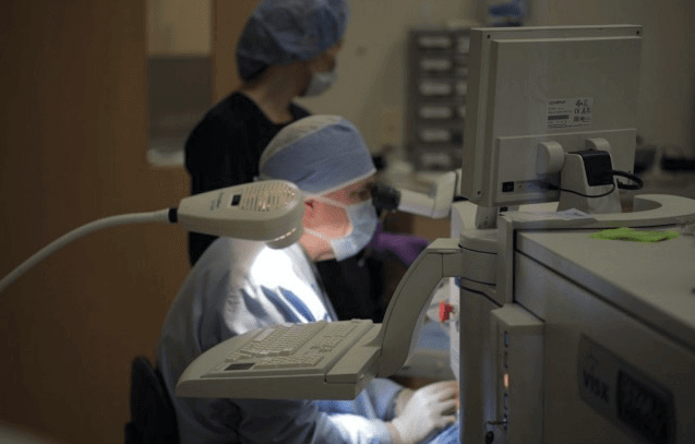
Surgery is usually done on an outpatient basis. You may be asked to skip breakfast, depending on the time of your surgery. When you arrive for surgery, you will be given eyedrops and perhaps a sedative to help you relax. A local anesthetic will numb the eye area. Your eye will be kept open by a special instrument. You may see light and movement, but you will not be able to see the surgery while it is happening.
The skin around your eye will be thoroughly cleansed, and sterile coverings will be placed around your head.
Under an operating microscope, a small incision is made in your eye. Microsurgical instruments are used to break apart and suction the cloudy lens from your eye. The back membrane of the lens (called the posterior capsule) is left in place. An intraocular lens implant will be placed inside your eye to replace the natural lens that was removed. The incision is then closed. When stitches are used, they usually do not need to be removed. When the surgery is complete, the doctor will often place a shield over your eye.
After a short stay in the outpatient recovery area, you will be ready to go home.
Following surgery
You will need to:
Ask your ophthalmologist if you should continue to take your usual medications.
- use the eyedrops as prescribed
- be careful not to rub or press on your eye
- use over the counter pain medicine if necessary
- avoid very strenuous activities until the eye has healed
- continue normal daily activities and moderate exercise
- ask your doctor when you can begin driving
- wear eyeglasses or eye shield as advised by your doctor
